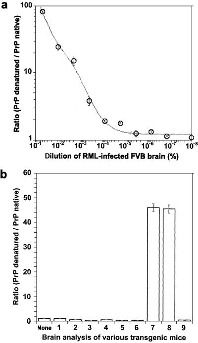
Figure 3.
CDI analysis of Tg(MoPrP,Q167R) mice. (a) Calibration curve performed on brain extracts of inoculated FVB mice. The europium-labeled HuM-D13 Fab was used for the detection of PrP. (b) CDI analysis of various brain homogenates. None, uninoculated wt FVB mice; 1 and 2, two prion-inoculated Tg(MoPrP,Q167R)Prnp0/0 mice killed after 550 days; 3 and 4, two uninoculated Tg(MoPrP,Q167R)Prnp0/0 mice killed after 420 days; 5 and 6, prion-inoculated FVB/Prnp0/0 mice killed after 420 days; 7 and 8, prion-inoculated Tg(MoPrP,Q167R)Prnp+/+ mice killed after 300 days; 9, an uninoculated Tg(MoPrP,Q167R)Prnp+/+ mouse killed after 150 days. Data points and bars are average ± SEM obtained from three independent measurements.

