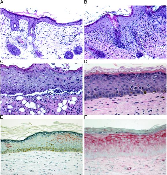
Figure 1.
(A–D) Topical titration of colchicine treatment demonstrates dose-dependent effects on mouse and human skin (2 wk). Shown are hematoxylin/eosin staining of mouse skin treated with vehicle control cream (×20) (A); colchicine (200 μg/g) treatment with increased numbers of KC blocked in mitosis (×20) (B); and colchicine (500 μg/g) treatment with cellular necrosis, severe disruption of the epidermal architecture, and ulceration (×20) (C). (D) Hematoxylin/eosin staining of human skin grafts treated with colchicine (200 μg/g) demonstrating KC blocked in mitosis but no disruption of the epidermal architecture (×40). (E and F) Immunohistostaining (alkaline phosphatase) for MDR1 (P-glycoprotein) expression in MDR+KC grafts (×40). (E) No colchicine treatment, 4 wk after grafting. (F) colchicine treatment (200 μg/g), 7 wk after grafting.

