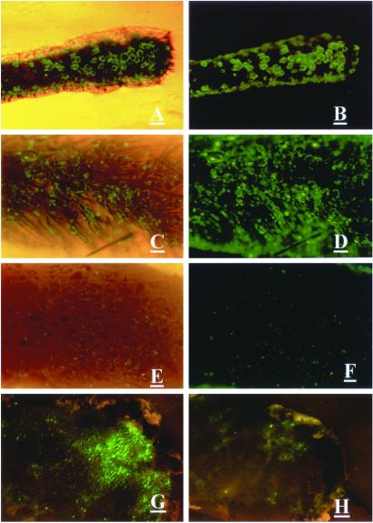
Figure 1.
Effect of collagenase treatment on adenoviral GFP transduction of hair follicles in skin. (A–D) Histocultured mouse skin treated with collagenase and transduced with adenoviral GFP. (A and C) Bright field and fluorescence. (B and D) Fluorescence. (E and F) Untreated histocultured skin transduced with adenoviral GFP. (Magnification = ×40; bar = 0.2 mm.) (G) Grafted skin, transduced with adenoviral GFP after collagenase treatment ex vivo. (H) Untreated grafted skin, transduced with adenoviral GFP. (Bar = 1 mm.) (E) Bright field and fluorescence. (F) Fluorescence. (A–F) The view from the undersurface. (G and H) The view from the surface.

