Figure 3.
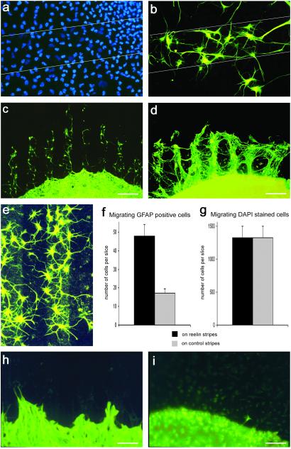
Response of GFAP-positive cells and processes in the stripe-choice assay. (a) All migrating cells, not distinguishing between different cell types, on a coated nucleopore membrane, visualized with the fluorescent nuclear stain DAPI (slice from P4, 14 DIV). The borders of a Reelin-coated stripe are indicated by lines. The DAPI-stained cells appear to be distributed evenly, without a preference for either the Reelin-coated stripe or the adjacent control stripes. (Stripe width, 90 μm.) (b) Same detail as in a after change of fluorescence filters. A minority of the migrating cells stained for GFAP show a clear preference for the Reelin stripe. (c) The arrangement of GFAP-immunostained cells strikingly reflects the striped pattern of the coated nucleopore matrix. GFAP-positive cells and their processes are located almost exclusively on the Reelin-coated stripes (slice from P4, 14 DIV). (Bar, 200 μm.) (d) GFAP-positive cells and fibers migrating out of a reeler mouse hippocampal slice culture (slice from P6, 14 DIV). The outgrowth of GFAP-positive glial processes is much more robust than that from wild-type hippocampal cultures (c). (Bar, 200 μm.) (e) GFAP-positive cells are preferentially located on a Reelin stripe. Reelin coating is visualized by immunostaining with an mAb against Reelin and a green fluorescent secondary antibody (slice from P6, 14 DIV). (Stripe width, 90 μm.) (f) Diagram representing the number of GFAP-positive cells on Reelin-containing stripes and control stripes. (Bar, SEM; n = 20 slice cultures from P4, 14 DIV.) (g) Diagram representing the number of DAPI-stained cells on Reelin-containing stripes and control stripes. (Bar, SEM; n = 8 slice cultures from P4, 14 DIV.) Note that similar numbers of DAPI-stained cells were found to migrate on Reelin-coated stripes and control stripes. The preferred adhesion of the few GFAP-positive cells (f) is masked by the large number of other cells, most likely microglial cells. (h) GFAP-positive cells and processes from hippocampal slice cultures (P4, 14 DIV) of scrambler mice do not show a preference for either the Reelin-coated stripes or the control stripes. (Bar, 90 μm.) (i) Only occasionally were GFAP-positive migrating cells or processes observed on the stripe matrix when hippocampal slices (P4, 14 DIV) from β1 integrin-deficient mice were cultured. No preference for Reelin stripes or control stripes was observed. (Bar, 90 μm.)

