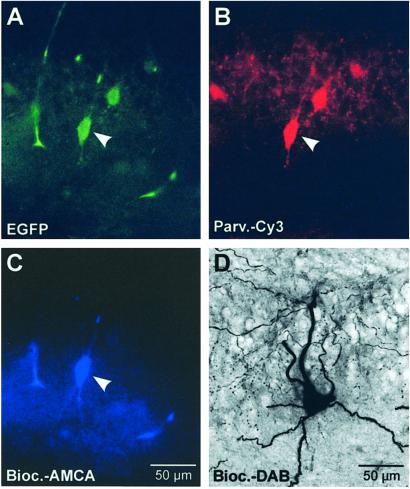
Figure 1.
Identification of parvalbumin-expressing BCs in slices of transgenic mice. (A) EGFP labeling, (B) parvalbumin immunoreactivity probed with anti-parvalbumin antibody and Cy3-conjugated secondary antibody, and (C) biocytin labeling with AMCA-conjugated avidin. Images were taken from the same cell in the CA1 pyramidal cell layer (arrowheads). (D) Light-microscopic image of a biocytin-labeled interneuron (EGFP-positive) in the CA3 region visualized using DAB as a chromogen. Note axonal arborization in the pyramidal cell layer.

