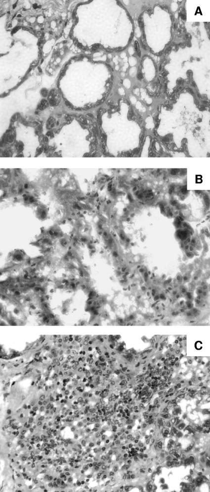
FIG. 2.
Hematoxylin-eosin-stained sections of mammary glands after challenge with 105 CFU of S. aureus. Depicted are representative sections exhibiting minor or no changes induced 4 days after challenge with Reynolds (CP−) (A), mild inflammation following challenge with Reynolds (CP8) (B), and a massive inflammatory response with marked PMN cell infiltration induced by Reynolds (CP5) (C).

