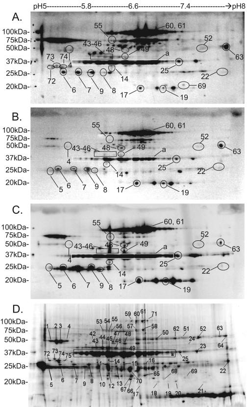
FIG. 3.
Identification by immunoblotting of antigenic A. marginale outer membrane proteins separated by 2D electrophoresis. Outer membrane proteins were separated by 2D electrophoresis. One gel was stained with SYPRO Ruby for total protein detection (D), while proteins from three other gels were transferred to nitrocellulose membranes. Membranes were probed with immune sera diluted 1:200 from 04B90 (A), 04B91 (B), and 04B92 (C), and binding was detected by HRP-conjugated anti-bovine IgG secondary antibody followed by a chemiluminescent substrate. To identify spots of interest, images from the SYPRO Ruby gel and the immunoblots were overlaid. Immunoreactive spots are labeled with numbers or blocks. Block a represents protein spots 26 to 41, identified as MSP2. Spots 60 and 61 represent MSP3, and spot 17 represents MSP5. The remaining immunogenic protein spots have been labeled on the immunoblots. Spots 12 and 70 were not immunoreactive but were used as two of several reference spots to determine reproducibility of outer membrane separated 2D gels. Spots 6, 63, and 17 were used to align 2D immunoblots. See Table 1 for a complete list of identified proteins.

