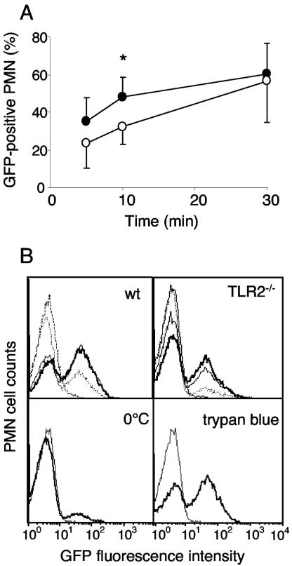
FIG. 2.
(A) Time course of phagocytosis of GFP-C5017 S. pneumoniae by purified mouse blood PMN analyzed by FACS and expressed as the percentage of PMN containing GFP bacteria after 5, 10, and 30 min. WT PMN (black circles) and TLR2−/− PMN (open circles). Flow cytometry acquisition was gated on PMN cells based on their forward scatter/side scatter and viability (negative propidium iodide staining). Mean values ± standard deviations from five independent experiments. Paired samples of WT and TLR2−/− cells were compared with the nonparametric analysis of variance (*, P < 0.05). (B) Fluorescence histograms of GFP-positive PMN during phagocytosis. PMN incubated 5, 10, and 30 min (dotted line, thin solid line and thick solid line, respectively) at 37°C (upper histograms) and WT PMN incubated 10 min at 0°C (lower left histogram) or 10 min at 37°C adding trypan blue to quench extracellular GFP-S. pneumoniae (lower right histogram) are shown. C5017 WT control (dashed line in the upper histograms and thin solid line in the lower histograms) is also shown. One representative out of three experiments is shown.

