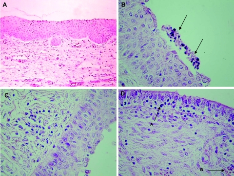
FIG. 3.
Photomicrographs of vagina, uterine corpus, and uterine horn tissue sections. All sections were stained with hematoxylin and eosin and visualized microscopically (magnification, ×400). (A) Typical uninfected vaginal morphology. (B) Inflammation of the vagina of a 468-infected gilt of group A. Note the presence of apoptotic-like cells above the epithelial layer (arrows) as well as few cells with intracellular edema in the epithelial layer. (C) Cervix of a Bour-infected gilt of group B. Note the diffuse infiltration of lymphocytes. (D) Uterine horn of a 468-infected gilt. Note the strongly folded epithelial layer with transmigrating lymphocytes and neutrophils (arrow A). The lamina propria shows focal infiltration of inflammatory cells and congestion of the blood vessels (arrow B).

