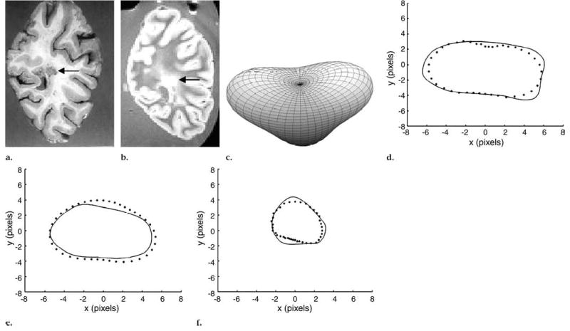
Figure 5.

Periventricular lesion (arrow) that extended into three coronal MR sections (550/20, field of view of 24 cm, and matrix size of 256 × 256 pixels). (a) Pathologic slice, (b) one of three MR sections, and (c) approximated 3D lesion shape obtained with SH method show pyramidal shape of lesion. (d–f) Segmented axial contours (solid line) versus corresponding contours from 3D-reconstructed shape with SH method (dotted line).
