
Figure 1.
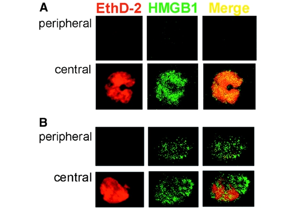
Immunofluorescence analysis of HMGB1 in resting and LPS-activated monocytes. Monocytes, freshly isolated (A) or cultured for 18 h with LPS (B), were fixed, permeabilized, stained with ethidium homodimer-2 (EthD-2, red channel) and anti-HMGB1 antibody (HMGB1, green channel) and analyzed by confocal microscopy. Two sections of the same cells (peripheral and central) are shown, which allows the nuclear (central) and cytoplasmic (peripheral, absent at time 0, and evident after 18 h of activation) staining to be appreciated. The merged images (Merge) verify the almost complete colocalization of HMGB1 and EthD-2 at time 0 (A, yellow) which decreases after LPS activation (B).