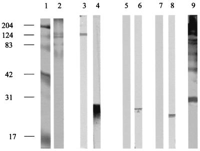
FIG. 1.
Western blotting profiles of MAbs against R. prowazekii. Lane 1, molecular size markers (numbers on the left are in kilodaltons); lane 2, Coomassie brilliant blue-stained, native R. prowazekii antigen (SDS-PAGE); lanes 3, 5, and 7, MAb P11A12; lanes 4, 6, and 8, MAb P9G1; lane 9, polyclonal antisera against native antigen (lanes 3, 4, and 9), heated antigen (lanes 5 and 6), and proteinase K-digested antigen (lanes 7 and 8).

