
Figure 5.
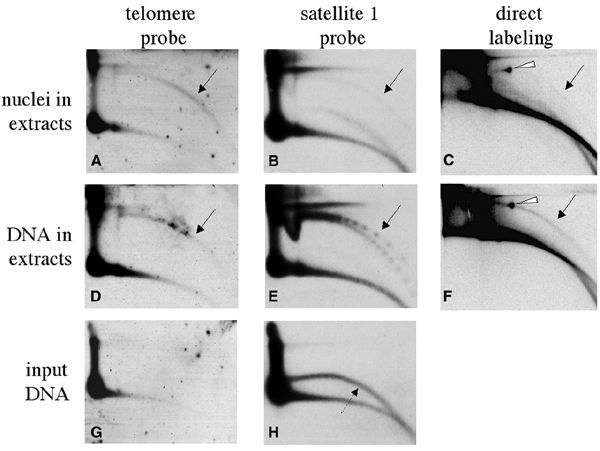
In vitro formation of tel-eccDNA from sperm nuclei and from naked sperm DNA. Sperm nuclei (270 ng of DNA) (A–C) or 300 ng of purified naked sperm DNA (D–F) were incubated for 2 h in 100 μl of low-speed Xenopus egg extract in the presence (C and F) or absence (A, B, D and E) of 50 μCi of [α-32P]dCTP. Following incubation, the DNA was extracted and analyzed by a 2D gel as well as non-incubated naked sperm DNA (G and H). The gel was hybridized with telomeric probe (A, D and G) and with satellite 1 probe (B, E and H). The gel that contained the radiolabeled samples was exposed to autoradiography (C and F). The solid arrows point to arcs of relaxed circles. In (E), a series of supercoiled multimers is also identified. The dashed arrow points to single-stranded DNA in the preparation of naked sperm DNA, which was previously shown to be irrelevant to eccDNA formation, as its degradation by S1 nuclease does not alter the production of eccDNA (Cohen and Méchali, 2001). The white arrowheads point to mtDNA that is present in the extract and was also labeled.