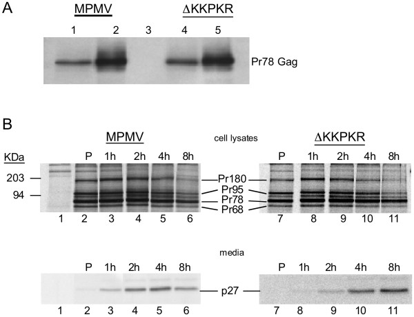
Figure 2.
(A) Western Blot analysis of intracellular procapsid assembly using Gag fractionation techniques. 48 hrs post transfection, COS-1 cells were lysed with and fractionated over a 20% sucrose cushion to separate assembled procapsids from unassembled Gag proteins. Pr78 in the fractionated samples were detected by western blot using rabbit anti-Pr78 antibodies. Soluble wild-type Pr78 (lane 1); pelletable wild-type Pr78 (lane 2); untransfected (lane 3); soluble ΔKKPKR Pr78 (lane 4); pelletable ΔKKPKR Pr78 (lane 5). (B) Virus release kinetics. Transfected COS-1 cells pulsed labeled with [35S] methionine-cysteine for 30 minutes and chased for 0, 1, 2, 4, and 8 hours. Untransfected (lane 1); pSARM-4 (lanes 2–6); ΔKKRKR (lanes 7–11). Medium was collected and cells were lysed at the appropriate times with 1× Buffer A. Cellular lysates and medium were adjusted to 1× lysis buffer B. Viral proteins were immunoprecipitated from all samples using rabbit anti-Pr78 antibodies and separated by SDS-PAGE (12% acrylamyde) and detected by phospor-imaging.

