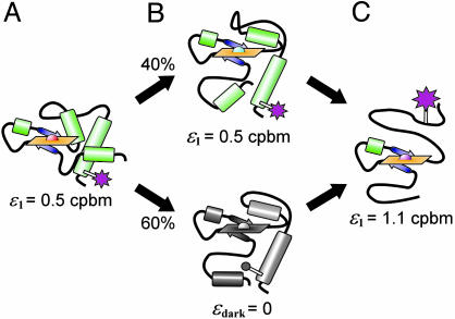
Fig. 5.
Structural model for the pH-dependent conformations of TMR-cytochrome c resolved by PCH (see Fig. 4). (A) At pH 7.0, the fluorophore (magenta) has a brightness of ε1 = 0.5 cpbm. (B) During the alkaline transition, 60% of species 1 enters a dark state and PCH resolves two discrete conformations. (C) As the protein unfolds, the fluorophore either brightens (B Upper) or leaves the dark state (B Lower) until ε1 reaches a maximum of 1.1 cpbm.

