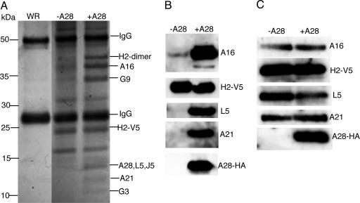
Fig. 1.
Identification of viral proteins associated with a putative entry–fusion complex. (A) Cells were infected with vA28i-HA/H2-V5 in the presence (+A28) or absence (-A28) of IPTG or with control VACV (WR). Detergent-extracted proteins from a viral membrane-enriched fraction were bound to V5 antibody covalently linked to agarose beads and analyzed by SDS/PAGE. Protein bands stained with Coomassie blue dye are shown with the positions of mass markers on the left. Bands were excised from the sample with IPTG (+A28) and analyzed by mass spectrometry; the identified proteins are indicated on the right. (B) Immunoaffinity-purified samples with (+A28) and without (-A28) IPTG used in A were analyzed by Western blotting with antibodies to A16, A21, HA, L5, and V5 as indicated. (C) Virions from cells infected with vA28HAi/H2-V5 in the presence (+A28) or absence (-A28) of IPTG were purified by sucrose gradient sedimentation and analyzed by Western blotting with the antibodies used in B.

