
Figure 2.
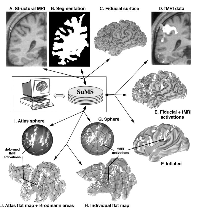
Illustrative data set taken through the processing sequence illustrated in Figure 1▶. A, Coronal slice through a structural MRI volume of human left hemisphere. B, Cortical segmentation through the same coronal slice, generated using the SureFit method. C, The fiducial surface generated from this segmentation. D, Functional MRI (fMRI) data overlaid on the same structural MRI data shown in A, generated in a behavioral paradigm involving eye movements.23 E, The fMRI data painted on the cortical surface and displayed in white and light shades. F, The same fMRI data displayed on an inflated surface, with cortical geography (sulcal regions) shown in darker shades. G, Spherical map that shows fMRI data, cortical geography, and latitude-longitude isocontours. H, The same data shown on a cortical flat map. I, Spherical map of the Visible Man atlas, with fMRI data deformed to the atlas. J,.Flat map of the Visible Man atlas that includes cortical geography, deformed fMRI data projected from the sphere to the flat map, and boundaries of Brodmann's architectonic areas as mapped by Drury et al.20 The data for the individual case can be downloaded from SuMS via a hyperlink connection to http://stp.wustl.edu/sums/sums.cgi?specfile=Demo.L.full.jamia.Fig2.spec.