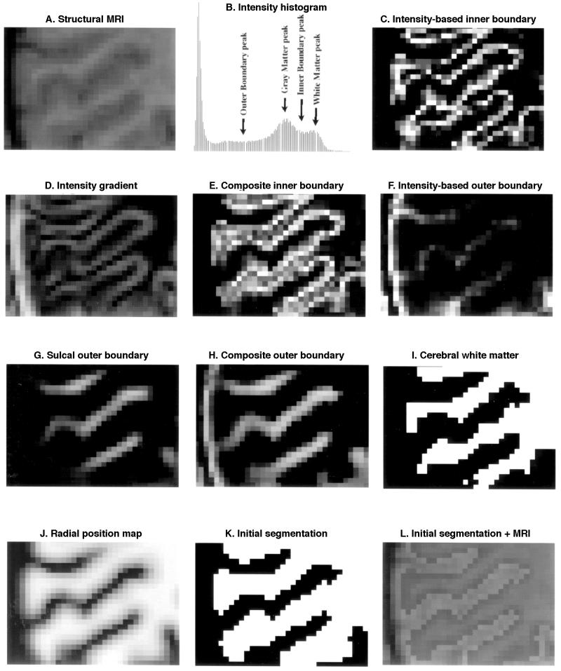
(Opposite) Stages of cortical segmentation in SureFit.
A, Coronal slice through occipital cortex in a human structural MRI volume.
B, Intensity histogram for the image volume shown in
A. Arrows indicate values for parameters used to generate intensity-based probabilistic maps of cortical regions or boundaries. C, An intensity-based probabilistic map of the inner boundary. This is based on a Gaussian intensity transformation:
and
where
I(n) is the intensity value for the th voxel,
Ipeak is an estimate of the most likely intensity value for the tissue type or boundary, and the standard deviations σ
low and σ
high are related to the noisiness of the image data.
D, Map of the magnitude of the intensity gradient.
E, The composite inner boundary map. One component of this composite map is the intensity-based map of the inner boundary
(C). Another component is derived by determining where the intensity gradient is intermediate in magnitude (made explicitly by a Gaussian intensity transformation analogous to that in
C) and pointed opposite to the gradient of a probabilistic map of matter (not shown; also based on a Gaussian intensity transformation). A third component selectively emphasizes regions near the crowns of gyri (where the underlying white matter is notably thin) by testing for regions containing two gradient-based inner-boundary domains that are in close proximity but pointed in opposite directions.
F, Intensity-based map of the outer boundary.
G, Map of the outer boundary in sulcal regions, based on evidence for two inner boundaries that are pointed in opposite directions and are each displaced about one cortical thickness (3 mm for human cortex) from the voxel being tested.
H, The composite outer boundary map, derived from the intensity-based outer boundary map
(F), the sulcal outer boundary
(G), and gradient-based cues analogous to those used for the inner boundary.
I, Binary map of cerebral white matter, based on thresholding the intensity volume and removing various noncerebral structures.
J, A map of positions along the radial axis, generated by blurring both the inner and outer boundary maps, normalizing the output by dividing the difference by the sum at each voxel, and assigning a maximal value to contained in the interior of cerebral white matter (i.e., in an eroded version of the image shown in
I).
K, The initial segmented volume obtained by thresholding the radial position map.
L, The initial segmented volume superimposed on the original intensity volume.