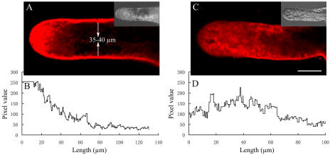
Figure 2.
Confocal images of FM4-64 staining in pollen tubes of P. meyeri. A, A median focal plane confocal optical section, showing a typical FM4-64 staining pattern in a growing pollen tube; the bright field at one-third size appears as an insert. B, Pixel values along a central transect through the fluorescence image in A. C, A median focal plane confocal optical section, showing a dispersed and disrupted FM4-64 staining pattern in BFA-treated (5 μg mL−1) pollen tube; the bright field at one-third size appears as an insert. D, Pixel values along a central transect through the fluorescence image in C.

