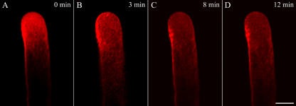
Figure 3.
Time course of disruption in a normal typical FM4-64 staining induced by 5 μg mL−1 BFA. BFA was applied directly to growing pollen tubes on thin gel layers in 70 μL of 115% to 120% liquid medium. A, A typical FM4-64 distribution pattern in a normal growing pollen tube of P. meyeri. B, The changes of FM4-64 staining at 3 min after the addition of BFA, showing the FM4-64 fluorescence tended to be scattered. C and D, The changed FM4-64 staining distribution with increasing time, showing FM4-64 fluorescence became more dispersed and almost distributed in the whole pollen tube after 12 min of BFA treatment. Bar = 25 μm.

