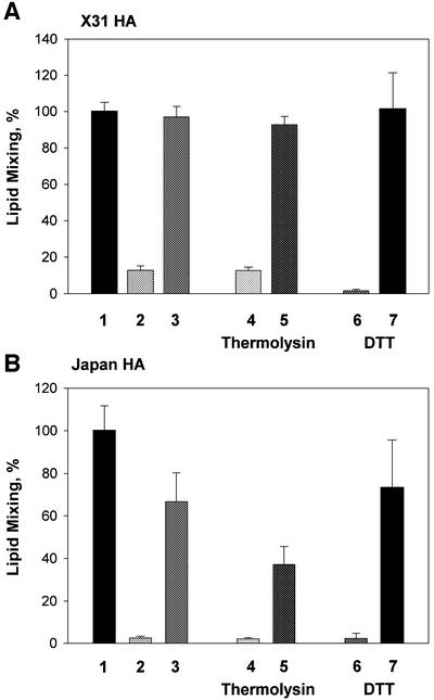
Fig. 1. Reversible conformations of low-pH-activated HA identified by means of a fusion inactivation assay. X31 (A) or Japan HA-cells (B) at 22°C were first treated with a 5 min pH 4.9 pulse (‘AP’) in the absence of target membrane. Then, the cells were incubated at neutral pH for different times. Finally, the cells were incubated with RBCs for 15 min and, after removal of unbound RBCs, the second (‘fusion-triggering’) low-pH pulse, FTP [a 2 min pH 4.9 pulse in (A) and a 5 min pH 5.2 pulse in (B)] was applied. Here and in the experiments reported in the following figures, the final extents of lipid mixing were measured by fluorescence microscopy. The total time intervals between the activating and fusion-triggering pulses were 15 min (bars 2) and 45 min (bars 3–7). Bars 4 and 5, thermolysin (25 µg/ml), and bars 6 and 7, DTT (10 mM) were applied for either the first (bars 4 and 6, respectively) or the last (bars 5 and 7, respectively) 5 min of incubation between the activating and fusion-triggering pulses. Fusion extents were normalized to those in the control experiments, in which the FTP [a 2 min pH 4.9 pulse in (A) and a 5 min pH 5.2 pulse in (B)] was applied to the HA-cells with bound RBCs untreated with an AP. Fusion extents of 83.9 and 63.5% were taken as 100% in bars A1 and B1, respectively.

An official website of the United States government
Here's how you know
Official websites use .gov
A
.gov website belongs to an official
government organization in the United States.
Secure .gov websites use HTTPS
A lock (
) or https:// means you've safely
connected to the .gov website. Share sensitive
information only on official, secure websites.
