Figure 4.
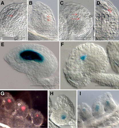
GUS expression patterns of the promoter∷GUS fusion with female gametophyte genes in early staged ovule. A to D, Ovule of wild-type plant at FG1 (A), FG2 (B), FG3 (C), and FG4 (D). A, The arrow points the nucleus of functional megaspore. B, Each arrow points a micropylar nucleus or chalazal nucleus within embryo sac. C, The two red arrows point the micropylar nuclei or chalazal nucleus, and a black arrow points a large central vacuole (cv). D, Embryo sac has two micropylar nuclei (red arrows), two chalazal nuclei (black arrows), and a large central vacuole (cv). E, The GUS activity of the promoter∷GUS fusion with At1g26795 was detected in embryo sac at FG4. F, GUS expression of the promoter∷GUS fusion with At1g36340 was detected in the closest nucleus to the chalazal end of the embryo sac at FG4. G, GUS activity of the promoter∷GUS fusion with At2g20070 was detected in dividing nuclei of the FG1 and FG2 embryo sac stages under the dark-field microscope. H, GUS expression of the promoter∷GUS fusion with At4g22050 was observed in chalazal end of embryo sac at FG4. I, GUS activity of the promoter∷GUS fusion with At5g40260 was detected in embryo sac at FG2. Scale bars represent 50 μm.

