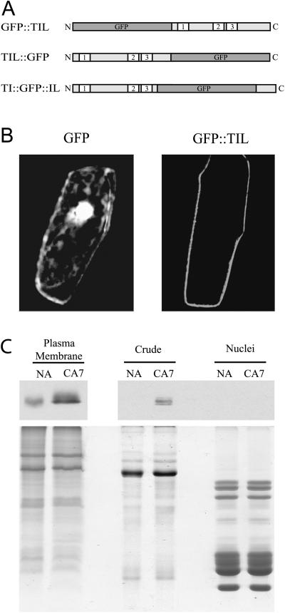
Figure 2.
Cellular localization of the plant TIL lipocalins. A, Schematic representation of GFP fusions used in the transient expression experiments. N and C are the amino and carboxy termini of the proteins, respectively; 1, 2, and 3 indicate the three SCRs. B, Transient expression assays of GFP-TIL fusions. Plasmids carrying the fusions were transformed into onion epidermal cells by microprojectile bombardment. Confocal images of GFP fluorescence were captured 20 h after transformation. Only the GFP∷AtTIL data are shown since the three constructs gave the same fluorescence pattern. The color figure is shown in Supplemental Figure 6. C, Biochemical fractionation analysis. Wheat protein extracts were prepared and subjected to SDS-PAGE and western-blot analyses. Top, Western-blot results obtained using the anti-TaTIL antibody (1/25,000, 10-s exposure for the PM fractions; 1/2,500, 5-min exposure for the other fractions). Bottom, Coomassie Brilliant Blue-stained gel showing the quality of the preparations. Typical protein patterns are observed for each fraction. NA, Nonacclimated plants grown for 7 d; CA7, plants grown for 7 d at 24°C, then cold acclimated at 4°C for 7 d.

