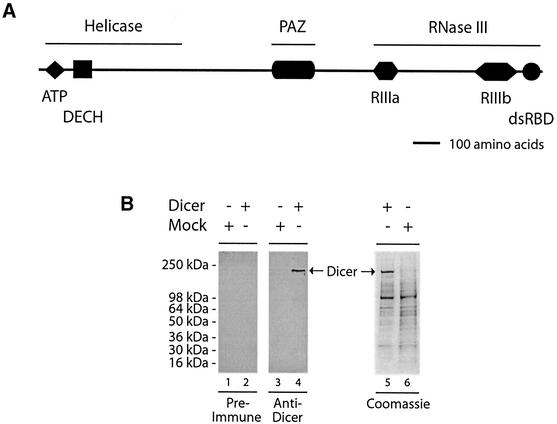
Fig. 1. Domain structure and expression of human Dicer. (A) Schematic illustration of the domain structure of the human Dicer protein. (B) Polyhistidine-tagged human Dicer protein was expressed in a baculovirus-based system, partially purified by Ni2+-affinity chromatography, and analyzed by SDS–PAGE followed by western blotting with anti-Dicer antibody or pre-immune serum, or Coomassie Blue staining. A preparation from mock-infected Sf9 cells was used as a control.

An official website of the United States government
Here's how you know
Official websites use .gov
A
.gov website belongs to an official
government organization in the United States.
Secure .gov websites use HTTPS
A lock (
) or https:// means you've safely
connected to the .gov website. Share sensitive
information only on official, secure websites.
