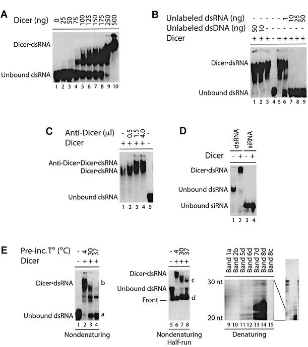
Fig. 2. Dicer interacts with dsRNA in EMSA. (A) 32P-labeled dsRNA was incubated in the absence or presence of Dicer (no MgCl2 added), (B) without or with unlabeled dsRNA or dsDNA poly(dA·dT), or (C) without or with anti-Dicer antibody (0.5–4.0 µl; no MgCl2 added). (D) 32P- labeled CLP 23 nt siRNA or 5LO dsRNA (100 bp) was incubated in the absence or presence of Dicer. (E) Left panel, 32P-labeled dsRNA was incubated in the absence or presence of Dicer, at the indicated temperatures for 1 h prior to EMSA analysis. Middle panel, same as left panel, but run half way. Dicer·dsRNA complex formation was analyzed by non-denaturing PAGE and autoradiography. Right panel, RNA was extracted from the indicated bands and analyzed by denaturing PAGE and autoradiography. The 20–30 nt region is shown, with the full-length gel on the right.

An official website of the United States government
Here's how you know
Official websites use .gov
A
.gov website belongs to an official
government organization in the United States.
Secure .gov websites use HTTPS
A lock (
) or https:// means you've safely
connected to the .gov website. Share sensitive
information only on official, secure websites.
