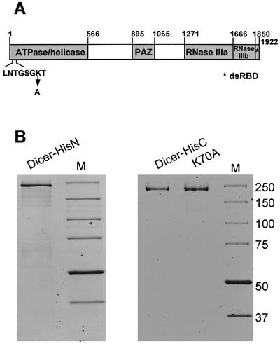
Fig. 1. Schematic structure of Dicer and SDS–PAGE analysis of recombinant Dicer preparations. (A) Domains of human Dicer. Amino acid positions corresponding to domain borders, and a P-loop sequence with the K70A mutation are indicated. (B) Dicer-HisN (1 µg), Dicer-HisC (0.5 µg) and Dicer-HisC P-loop mutant K70A (1 µg) were analysed by 8% SDS–PAGE. Proteins were stained with GelCode Blue Stain Reagent (Pierce). Lanes M, protein size markers in kDa (BenchMark Protein Ladder, Invitrogen).

An official website of the United States government
Here's how you know
Official websites use .gov
A
.gov website belongs to an official
government organization in the United States.
Secure .gov websites use HTTPS
A lock (
) or https:// means you've safely
connected to the .gov website. Share sensitive
information only on official, secure websites.
