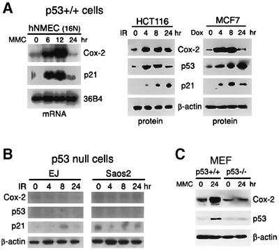
Fig. 2. Induction of Cox-2 in response to DNA damage. (A) Cox-2 expression in wild-type p53-containing cells treated with DNA-damaging agents. 16N hNMECs were exposed to 2.5 µg/ml MMC for 0, 6, 12 or 24 h. Total RNA was then extracted and analyzed by northern blotting. HCT116 and MCF-7 cells were exposed to 5 Gy of γ-irradiation (IR) or 0.3 µg/ml of doxorubicin (Dox), and total proteins were extracted at the indicated times after treatment and subjected to western blot analysis. (B) Cox-2 expression in p53-null cells in response to DNA damage. EJ (bladder cancer cell line) and Saos2 (osteosarcoma cell line) cells were γ-irradiated (5 Gy), and total protein lysates were isolated for western blot analysis. β-actin was used as a loading control. (C) p53+/+ or p53–/–MEFs were treated with 2.5 µg/ml MMC for 24 h. Total cell extracts were isolated for western blot analysis.

An official website of the United States government
Here's how you know
Official websites use .gov
A
.gov website belongs to an official
government organization in the United States.
Secure .gov websites use HTTPS
A lock (
) or https:// means you've safely
connected to the .gov website. Share sensitive
information only on official, secure websites.
