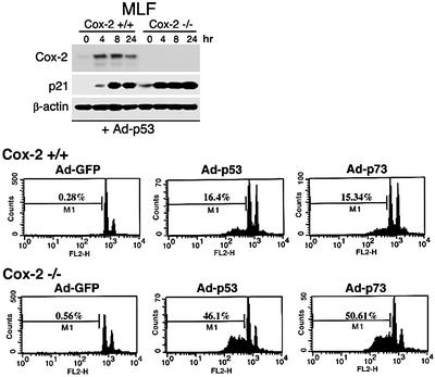
Fig. 4. p53-mediated apoptosis was enhanced in Cox-2-null cells. Cox-2+/+ or Cox-2–/– MLFs were infected with Ad–GFP, Ad-p53 or Ad-p73α for 24 h. Upper panel: a western blot analysis using lysates prepared from cells infected with Ad-p53 adenoviruses. Lower panels: the patterns of apoptosis analysis by FACScan. The M1 cell population represents apoptotic cells from each sample.

An official website of the United States government
Here's how you know
Official websites use .gov
A
.gov website belongs to an official
government organization in the United States.
Secure .gov websites use HTTPS
A lock (
) or https:// means you've safely
connected to the .gov website. Share sensitive
information only on official, secure websites.
