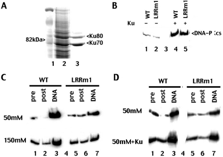
Figure 5.
The LRR contributes to DNA-PKcs's intrinsic affinity for DNA but not its interaction with Ku. (A) Whole cell extract from Sf9 cells infected with baculovirus expressing His-tagged Ku was incubated with Ni+ agarose beads. Beads were washed and analyzed for Ku expression by SDS–PAGE followed by Coomassie blue staining. (B) Immunoblotting of Ni+ agarose fractions of whole cell extracts (2 mg) from V3 transfectants expressing either wild-type DNA-PKcs (lanes1 and 4) or LRRm1 (lanes 2 and 5) incubated with whole cell lysate from either control virus infected-Sf9 cells or Ku-infected Sf9 cells as indicated. (C) Immunoblot analysis of DNA-cellulose fractions of extracts (500 µg) from V3 transfectants expressing wild-type (WT) DNA-PKcs (lane 3) or LRRm1 (lane 7) in either 50 mM salt (upper panel) or 150 mM salt (lower panel) as indicated. Lysates, pre-absorption (lanes 1 and 5) and post-absorption (lanes 2 and 6) represents 2% of the total lysates input. (D) Immunoblot analysis of DNA-cellulose fractions of extracts (500 mg) from V3 transfectants expressing wild-type (WT) DNA-PKcs (lane 3) or LRRm1 (lane 7) in either 50 mM salt (upper panel) or 50 mM salt suspplemented with partially purified Ku (expressed in Baculovirus) (lower panel) as indicated. Lysates, pre-absorption (lanes 1 and 5) and post-absorption (lanes 2 and 6) represents 2% of the total lysates input.

