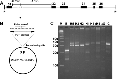
Figure 1.
(A) Map (to scale) of the palindromic region within a 3.8 kb EcoR1 fragment of the NF1 gene. Exons are numbered according to L05367 (see text). ‘R1’ denotes EcoR1 sites according to Southern blot data (27) and the reference human genome sequence (May 2004). (B) Cloning strategy. Oligos used for PCR flank the palindromic site. The PCR product is ligated into a commercial vector (see Materials and Methods). ‘X’ and ‘P’ refer to the Xbal and PvuII sites used in diagnostic digests. (C) PCR amplification of various DNA templates. Lane ‘M’, markers (Trackit 100 bp ladder, Invitrogen). ‘B’ is a PCR with Bac clone CTD-2370N5; ‘H1’ to ‘H5’ are with human genomic DNA from the indicated individuals. ‘C’ is with chimpanzee DNA. ‘φH4’ and ‘φG’ are from an aliquot of H4 and gorilla genomic DNA that had first been amplified with phi-29 polymerase. The H4 samples demonstrate reproducibility of the PCR as well as the fidelity of phi-29 amplification.

