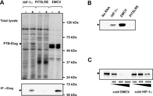
Figure 2.
PTB binds to the HIF-1α IRES. (A) Cytoplasmic extracts from HEK293T cells overexpressing E-tagged c-Raf (−) or PTB (+) were prepared as described in the ‘Materials and Methods’ section. After incubation with the RNA probes corresponding to the HIF-1α IRES, the EMCV IRES, or the PITSLRE IRES, the samples were UV-irradiated and treated with an RNase cocktail. One part of the sample was analyzed by SDS–PAGE (upper panel), the other was mixed with anti-E-tag antibody for immunoprecipitation of labeled E-tagged PTB. Bound proteins were analyzed by SDS–PAGE (lower panel). The arrows depict the position of crosslinked E-tagged PTB. (B) Biotinylated HIF-1α, EMCV and PITSLRE IRES RNAs bound to streptavidin beads were incubated with cytoplasmic extracts from parental HEK293T cells as described in the ‘Materials and Methods’ section. After extensive washing, RNA-bound proteins were analyzed by western blotting using anti-PTB antibodies. The arrow depicts the position of endogenous PTB. (C) Recombinant PTB was UV-crosslinked to radiolabeled HIF-1α IRES RNA in the absence or presence of a 10–500 molar excess of unlabeled EMCV IRES or HIF-1α IRES RNA. After RNA binding and UV-irradiation, samples were treated with an RNase cocktail and resolved by SDS–PAGE. The arrow depicts the position of crosslinked His-tagged PTB.

