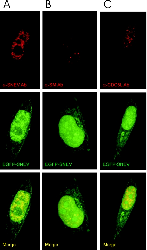
Figure 3.
SNEV associates in vivo with Sm proteins as well as with the splicing factor CDC5L, and fusion of SNEV to EGFP does not alter the cellular localization of SNEV. HeLa cells were grown, transfected with an EGFP–SNEV fusion construct, fixed and stained by indirect immunofluorescence. The images shown in the panels are representative optical sections from the respective deconvolved datasets. (A) GFP–SNEV expressing HeLa cells stained with anti-SNEV antibody 867. Red represents SNEV indirect staining, and green indicates EGFP–SNEV expression. (B) Cells were stained with the the anti-Sm protein monoclonal antibody Y12 (57). Red indicates Y12 staining as above, while green represents EGFP–SNEV expression. (C) Cells stained with anti-CDC5L antibodies. Red represents anti-CDC5L staining, while green shows EGFP–SNEV expression. In all the panels, yellow indicates co-localization of the two proteins. Note that the use of the fluorescently labelled protein results in a stronger signal from the fusion protein. This shows up as a small fraction of the label that does not co-localize.

