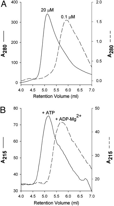
Fig. 1.
Dimerization assays of MalK by gel filtration. (A) Concentration-dependent dimerization in the absence of nucleotides. MalK at a 20 μM concentration (peak fraction) elutes at the expected position of a dimer, whereas at a concentration of 0.1 μM MalK elutes as a monomer. (B) Nucleotide-dependent dimerization at monomeric protein concentration. MalK stock at 1 μM concentration was mixed with 0.5 mM ATP (solid line) or 0.5 mM ADP and 10 mM MgCl2 (dashed line) and loaded to a size-exclusion column preequilibrated with buffer containing the corresponding nucleotide. The elution peak fraction contains MalK at ≈0.1 μM concentration. Note that in both A and B, for the purpose of comparison, the profiles do not use the same scale. Absorbance is shown in mAU. Figures were prepared with sigmaplot(Systat, Point Richmond, CA).

