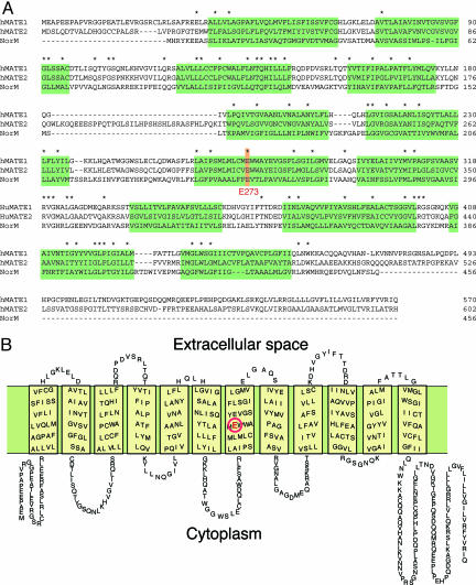
Fig. 2.
Amino acid sequences of hMATE1 and hMATE2. (A) The amino acid sequences of the proteins are aligned with that of NorM (14). Identical sequences are indicated by asterisks. Predicted transmembrane regions are shaded. (B) Putative secondary structure of hMATE1. A glutamate residue (E273) that is essential for the transport activity is shown in red (16).

