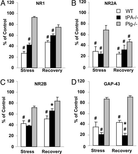
Fig. 2.
Quantification of stress-induced changes in NMDA receptor subunits and GAP-43. Animals (wild type, n = 4; tPA-/-, n = 4; plasminogen-/-, n = 3 for each time point) were subjected to chronic restraint stress, their hippocampi dissected, and the levels of individual NMDA receptor subunits and GAP-43 were determined by Western blotting. The membranes were stripped and reblotted for actin for loading control. Changes in the expression of NR1 (A), NR2A (B), NR2B (C), and GAP-43 (D) after chronic stress or stress followed by 10 days of recovery are shown and compared with the levels observed in stress-naïve control mice. Stress-induced changes were attenuated in tPA-/- mice and almost completely prevented in plasminogen-/- animals. For quantification, the background was digitally eliminated by using scion image, and optical densities of the bands were normalized to actin. *, P < 0.05; #, P < 0.001; * in D, P < 0.05 vs. plasminogen-/-.

