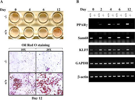
Figure 7. Ex Vivo Adipogenesis Analysis of Sam68−/− Mouse Embryonic Fibroblasts.
MEFs were isolated from mouse embryos at embryonic day 14.5. Equal number of MEFs from Sam68+/+ and Sam68−/− was plated on glass cover slips in 24 well-plates. Adipocyte differentiation was carried out at indicated times by the addition of complete media containing the pioglitazone.
(A) Cultures were fixed in 4% paraformaldehyde and stained with Oil Red O to detect the fat droplets stored in adipocytes and photographed (top). The cell images were magnified ×10 and ×20 as indicated.
(B) RT-PCR was carried out on total cellular RNA isolated after differentiation of the MEFs for day 0, 2, 4, 6, and 12. The DNA fragments were visualized on agarose gels stained with ethidium bromide. The expression of adipogenic markers C/EBPβ, C/EBPδ, PPARα, and KLF5 was examined as well as the expression of controls including Sam68, β-actin, and GAPDH.

