Figure 4.
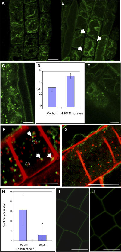
GFP-KOR1–Labeled Structures Lie on the Endocytic Pathway in Arabidopsis Root Cells, and Their Distribution Is Regulated during Isoxaben Treatment and Normal Cellular Development.
(A) and (B) Orthogonal projections from living root cells expressing GFP-KOR1 untreated (A) or treated with 4 × 10⁻⁹ M isoxaben for 1 h (B). Isoxaben treatment induces a redistribution of GFP-KOR1 compartments. Arrows indicate the dots at the tops of the cells. The strong background labeling corresponds to the tonoplast.
(C) and (E) Single optical sections of a root epidermal cell expressing GFP-KOR1 untreated (C) or treated with 4 × 10⁻⁹ M isoxaben for 1 h (E). Note the heterogeneous population of KOR1-labeled structures localized close to the cell surface in untreated root cells. A more homogeneous population of smaller compartments is detected in isoxaben-treated cells.
(D) Percentage of dots present at the tops of cells expressing GFP-KOR1 treated with or without 4 × 10⁻⁹ M isoxaben for 1 h. Note the redistribution of GFP-KOR1 toward the cortical region of cells upon isoxaben treatment. Error bars represent se.
(F) and (G) Single optical sections of a cell colabeled with FM4-64 and GFP-KOR1 in root cells untreated (F) or treated with 4 × 10⁻⁹ M isoxaben for 1 h (G). Note that some GFP-KOR1 colocalized with FM4-64 after 10 min of incubation (arrows) in untreated root cells. When not colocalized, they frequently appeared adjacent to each other (open circles). Note that upon isoxaben treatment, no colocalization was observed between GFP-KOR1 compartments and FM4-64–labeled structures, suggesting that isoxaben caused the redistribution of GFP-KOR1 away from early endosomes.
(H) Percentage of colocalization of FM4-64–labeled structures and GFP-KOR1 compartments in root cells from different growth stages. In smaller cells (10 μm), 15% of the GFP-KOR1 compartments colocalized with the early endosomal marker, compared with only 2.5% in the larger cells (cells between 50 and 70 μm). Error bars represent se.
(I) and (J) Single optical sections of a cell expressing the plasma membrane GFP-LTI6b fusion protein untreated (I) or treated with 4 × 10⁻⁹ M isoxaben for 1 h (J). No intracellular labeling was observed upon isoxaben treatments.
Bars = 10 μm.

