Figure 7.
Subcellular Localization of Fluorescent Protein-Tagged FRA8.
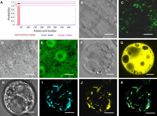
Fluorescent protein–tagged FRA8 was expressed in Arabidopsis plants and carrot protoplasts, and its subcellular location was examined with a laser confocal microscope. Bars = 9 μm in (B) to (E) and 14 μm in (F) to (K).
(A) FRA8 is a type II membrane protein as predicted by the TMHMM2.0 program. FRA8 is predicted to have a transmembrane helix between amino acid residues 21 to 38, a short N-terminal region located on the cytoplasmic side of the membrane (inside), and a long stretch of C-terminal region located on the noncytoplasmic side of the membrane (outside).
(B) and (C) Differential interference contrast (DIC) image (B) and the corresponding fluorescent signals (C) of Arabidopsis root epidermal cells expressing FRA8-GFP. Note that the FRA8-GFP signals show a punctate pattern.
(D) and (E) DIC image (D) and the corresponding fluorescent signals (E) of Arabidopsis root epidermal cells expressing GFP alone. Note the presence of GFP signals throughout the cytoplasm.
(F) and (G) DIC image (F) and the corresponding fluorescent signals (G) of a carrot cell expressing EYFP alone.
(H) to (K) DIC image (H) and the corresponding FRA8-ECFP signals (I), MUR4-EYFP signals (J), and a merged image (K) of a carrot cell expressing FRA8-ECFP and the Golgi-localized MUR4-EYFP. It is evident that the FRA8-ECFP signals are identical to MUR4-EYFP signals.

