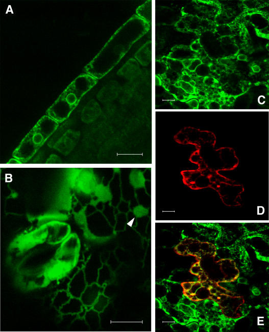
Figure 4.
Subcellular Localization of a Functional PHF1:GFP Fusion Protein.
(A) Confocal laser scanning micrographs of Pi-starved root cells expressing PHF1:GFP from the PHF1 promoter.
(B) Micrographs of Pi-starved leaf epidermal cells from a transgenic line harboring the 35S:PHF1:GFP construct, in which fluorescence highlights a polygonal network of ER tubules interspersed with small patches of ER lamellae, one of which is indicated with a white arrowhead.
(C) to (E) Micrographs of Pi-starved leaf epidermal cells from a transgenic line harboring the 35S:PHF1:GFP construct bombarded with a construct encoding an ER-located marker (35S:DsRed2:KDEL). The images shown correspond to PHF1:GFP (C), DsRed2:KDEL (D), or an overlay of the two (E).
Bars = 10 μm.

