Figure 5.
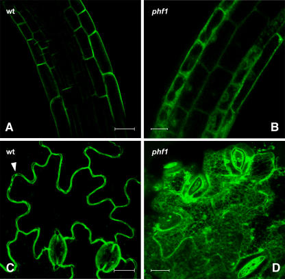
Subcellular Localization of PHT1;1:GFP in Wild-Type and phf1 Plants.
(A) and (B) Confocal laser scanning micrographs of Pi-starved transgenic wild-type (A) and phf1-1 (B) root cells expressing PHT1;1:GFP from the PHT1;1 promoter showing fluorescence associated with the plasma membrane and the ER, respectively.
(C) and (D) Micrographs of leaf epidermal cells from transgenic wild-type (C) and phf1-1 lines (D) harboring the 35S:PHT1;1:GFP construct. In (C), fluorescence highlights the plasma membrane, and a cell junction is indicated with a white arrowhead. By contrast, in (D), a reticulate structure is very evident.
Bars = 10 μm.

