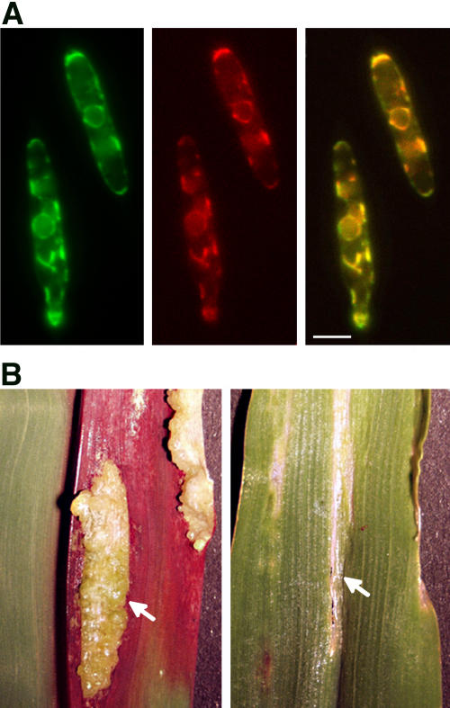
Figure 2.
Gas1-YFP Fusion Protein Localizes to the ER of U. maydis.
(A) Left panel: CFP fluorescence of CFP fusion protein that is targeted to the ER by a calreticulin signal peptide and a C-terminal ER retention signal (Wedlich-Söldner et al., 2002). Middle panel: YFP fluorescence of Gas-YFP fusion protein in the same cell. Since the Gas-YFP signal was faint and required long integration time, ER motility had to be stopped by brief formaldehyde treatment. The overall organization of the ER remained unaffected. Right panel: Merged images of left and middle panels showing colocalization of Gas1-YFP and ER-resident CFP. For clarity, signals emitted by CFP fluorescence were colored in green, while those emitted by YFP fluorescence were colored in red. Merged signals appear yellow. Bar = 3 μm.
(B) Pathogenicity symptoms on infected plants. Five-day-old maize seedlings were inoculated either with a mixture of haploid wild-type cells (FB1 × FB2) or with a mixture of Δgas1 mutant strains (HBU13 × HBU14). Ten days after infection, tumors are formed on plants infected with the wild type (left panel, arrow), while infection with Δgas1 strains leads to the formation of localized slightly necrotic areas on the leaf (right panel, arrow).

