Figure 5.
Δgas1 Strains Arrest Growth after Plant Penetration.
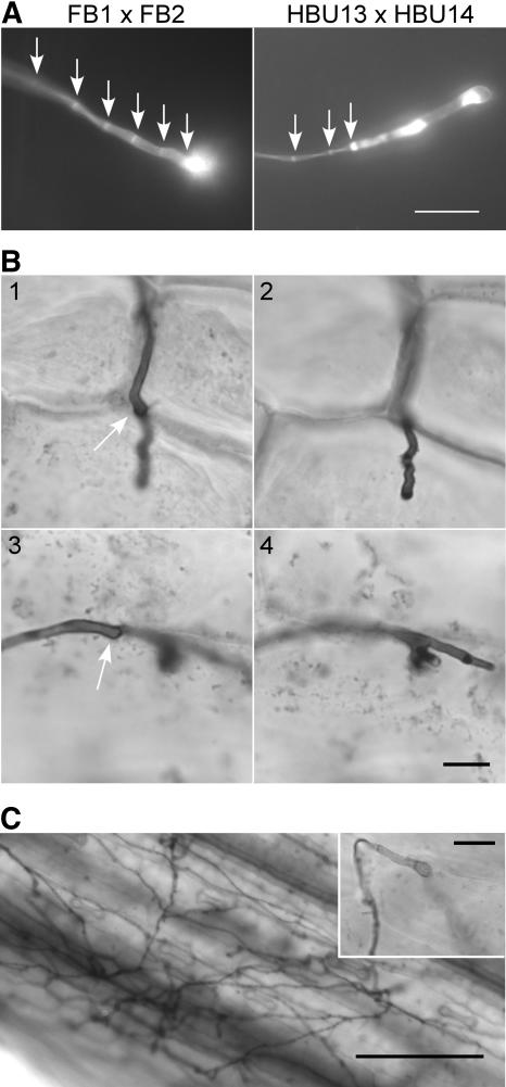
(A) Appressoria visualized by leaf staining with calcofluor 1 d after infection of young maize seedlings with a combination of wild-type (FB1 and FB2) or Δgas1 strains (HBU13 and HBU14). The wild-type appressoria show regular septation up to the appressorial tip (arrows), while in mutant appressoria, regular septation (arrows) does not continue up to the hyphal tip. The remaining elongated cell shows irregular distribution of calcofluor-stainable cell wall material. Bar = 10 μm.
(B) Δgas1 strains arrest growth after plant penetration. Maize seedlings were infected with a mixture of the Δgas1 strains HBU13 and HBU14. Two days after infection, leaf samples were stained with Chlorazole Black E and analyzed by light microscopy. Two different hyphae at their site of penetration are shown (1 and 3), also in a different focal plane (2 and 4), with the arrows indicating the appressorial tip on the plant surface. The invading hyphae grow into the plant cell but arrest growth (2) and may branch (4). Bar = 10 μm.
(C) Wild-type hyphae ramify extensively in the host leaf. Maize seedlings were infected with a mixture of FB1 and FB2. Two days after infection, leaf samples were stained with Chlorazole Black E and analyzed by light microscopy. The inset shows a typical wild-type appressorium with an invading hypha growing into the plant tissue out of focus. Bar = 100 μm (bar in inset = 10 μm).

