Figure 3.
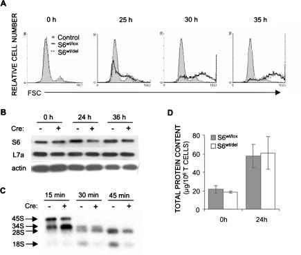
T-cell receptor-stimulated ribosome biogenesis is decreased in S6wt/del T cells, but their growth is normal. (A) FSC analysis of S6wt/lox and S6wt/del lymph node T cells stimulated with soluble 1 μg/mL anti-CD3 and 0.1 μg/mL anti-CD28 in vitro was used to determine their size. (B) The protein expression of ribosomal proteins S6 and L7a in lysates from unstimulated (0 h) and stimulated (24 and 36 h) lymph node S6wt/del and S6wt/lox T cells was analyzed by Western blot. Reprobing with antibodies to actin served as a loading control. (C) To gain insight into rRNA processing, S6wt/del and S6wt/lox T cells were stimulated for 20 h with TCR and CD28 antibodies, pulse-labeled with L-[methyl-3H]methionine for 30 min, and chased in nonradioactive medium for the indicated time. Total RNA normalized to equal cell number was resolved on a formaldehyde-agarose gel, transferred to a nylon membrane, and visualized by autoradiography. The positions of major precursors and 28S and 18S rRNA species are indicated on the left. The experiment is a representative of five independent experiments. (D) Total cellular protein content per 1 × 10⁶ S6wt/lox or S6wt/del lymph node T cells before stimulation and after 24 h of stimulation with antibodies against TCR and CD28. The experiment is a representative of six independent experiments.

