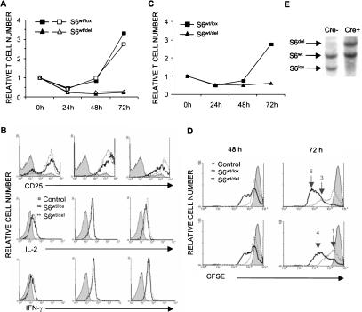
Figure 4.
Stimulation of S6wt/del T cells does not increase their cell number. (A) Relative number of S6wt/lox and S6wt/del T cells stimulated with either 0.1 μg (open symbols) or 1 μg/mL (closed symbols) of anti-CD3 and fixed concentration of anti-CD28 antibodies (0.1 μg/mL) during 72 h in vitro. The experiment is a representative of 15 independent experiments. (B) FACS analysis of expression levels of CD25, intracellular IL-2, and IFN-γ in S6wt/lox and S6wt/del lymph node T cells stimulated for the indicated period of time in vitro. (C) Relative number of S6wt/lox and S6wt/del lymph node T cells stimulated with soluble anti-CD3 antibodies and saturating concentration of IL-2 containing MLA-144 supernatant (1:2 dilution). The experiment is a representative of three independent experiments. (D) FACS analysis of lymph node T cells that were stimulated with different concentrations of anti-CD3 (1 μg/mL, upper panel; 0.1 μg/mL, lower panel) and anti-CD28 antibodies (0.1 μg/mL) and labeled with CFSE. The filled area represents CFSE labeling of T cells before they divided for the first time. The number of cell divisions is indicated above the peaks. (E) Southern blot analysis of genomic DNA from S6wt/lox and S6wt/del lymph node T-cell cultures at 72 h following stimulation.

