Figure 5.
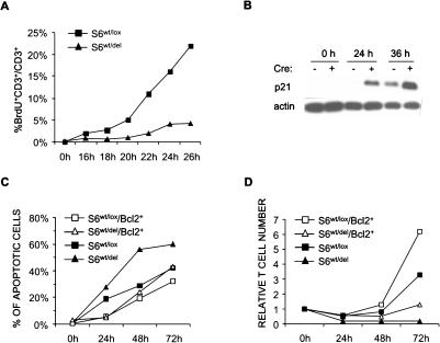
Defect in proliferation of S6wt/del T cells is due to p21-induced G1/S block. (A) BrdU incorporation into lymph node T cells stimulated with anti-CD3- and IL-2-containing MLA144 supernatant in vitro. The experiment is a representative of four independent experiments. (B) The protein expression of p21 cell cycle inhibitor in lysates from resting and stimulated lymph node S6wt/del and S6wt/lox T cells was analyzed by Western blot. Reprobing with antibodies to actin served as a loading control. (C) T cells of indicated genotypes were stimulated for the indicated period of time, labeled with propidium iodide, and analyzed by FACS. The percentage of apoptotic cells was determined by analyzing sub-G1 DNA content. The experiment is a representative of four independent experiments. (D) Relative number of S6wt/lox, S6wt/del, S6wt/lox/Bcl-2+, and S6wt/del/Bcl-2+ T cells stimulated with anti-CD3 and anti-CD28 antibodies for 24, 48, and 72 h. The experiment is a representative of three independent experiments.

