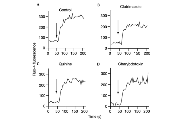
Figure 3.

The effect of IKCa1 inhibitors on Ca2+ uptake by CD4+ lymphocytes. Lymphocytes labelled with the Cychrome-conjugated anti-CD4 antibody were incubated with the Ca2+-indicator Fluo-4 AM, washed, and analysed by flow cytometry. Cells were stimulated with 1 μM calcimycin in the absence of inhibitor (A), or the presence of 10 μM clotrimazole (B), 0.5 mM quinine (C) or 200 nM charybdotoxin (D). An increase in Ca2+ in the cell is indicated by raised Fluo-4 fluorescence. Little or no effect of the IKCa1 inhibitors on Ca2+ influx was seen. Stimulation with calcimycin is indicated by arrows.
