
Figure 2.
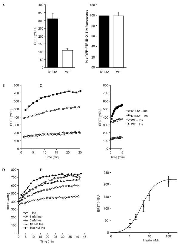
Dynamics of the interaction between the insulin receptor (IR) and protein-tyrosine phosphatase 1B (PTP1B) in intact living cells. (A) Basal bioluminescence resonance energy transfer (BRET) signal (left panel) and yellow fluorescent protein (YFP) fluorescence (right panel) in human embryonic kidney (HEK) cells co-expressing IR–Rluc—a fusion of the IR to Renilla luciferase (Rluc)—and YFP-tagged forms of either wild-type (WT) PTP1B or the D181A mutant form of PTP1B. Results are expressed as the mean ± s.e.m. (n = 5). (B) HEK cells co-expressing IR–Rluc and either YFP–PTP1B or YFP–PTP1B–D181A were incubated either in the absence of insulin, or in the presence of 100 nM insulin. (C) Early effect of insulin on the interaction between the IR and PTP1B. Results are representative of at least five independent experiments. (D) Dose-dependent effect of insulin on the interaction of the IR with the YFP–PTP1B–D181A substrate-trapping mutant. HEK cells were incubated either in the absence of insulin or the presence of different concentrations of insulin. (E) Dose-response curve of the insulin-induced BRET signal at t=20 min. Results are the mean ± s.e.m. of 4–7 independent experiments. Ins, insulin.