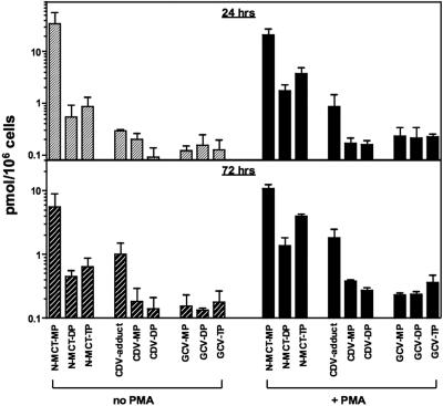
FIG. 3.
Intracellular phosphorylation profiles of N-MCT, CDV, and GCV in BCBL-1 cells with (+ PMA) and without PMA stimulation (no PMA). Shown are the levels of mono-, di-, and triphosphorylated metabolites of the test compounds analyzed at 24 h (top) and 72 h (bottom) postincubation. Of note, the phosphorylated metabolites of CDV were identified as CDV-monophosphate, CDV-DP (active metabolite), and a phosphate ester adduct of CDV (CDV-adduct) as previously described (28). The data shown are means ± standard deviations of two separate assays.

