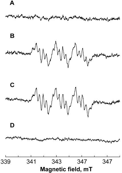
FIG. 2.
EPR spectra demonstrating formation of metronidazole anion radicals by the hydrogenosomes of low-resistance (strain TV 10-02 MR 5; traces A to C) and highly resistant (strain TV 10-02 MR 100; trace D) T. vaginalis strains in the presence of 43 mM metronidazole. (A) Absence of signal in the presence of pyruvate, ferredoxin, and CoA. (B) Signal generated in the presence of malate, ferredoxin, and NAD+ (malic enzyme activity). (C) Signal generated in the presence of NADH and ferredoxin (NDH activity). (D) Absence of signal in the presence of hydrogenosomes of highly resistant T. vaginalis, NADH, and ferredoxin (NDH activity). Instrument settings were as described for Fig. 1.

