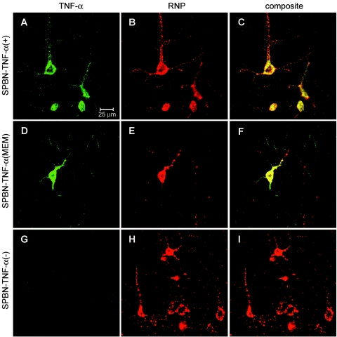
FIG. 5.
Expression of TNF-α (green) in cerebral neurons infected with SPBN-TNF-α(+) (A to C) or SPBN-TNF-α(MEM) (D to F), but not in SPBN-TNF-α(−)-infected neurons (G to I), on day 10 p.i. in the cortexes of TNF-α KO mice. High-power double-immunofluorescence laser scanning microscopic images of TNF-α (green) and rabies ribonucleocapsid (RNP, red) expression are shown.

